Cannabaceae is a small family of flowering plants, commonly known as the hemp family[1][2]. Here are the key details about Cannabaceae:
Classification and Taxonomy
Cannabaceae belongs to the order Rosales[1]. The family includes approximately 170 species grouped into about 11 genera[1][3]. The main genera in this family are:
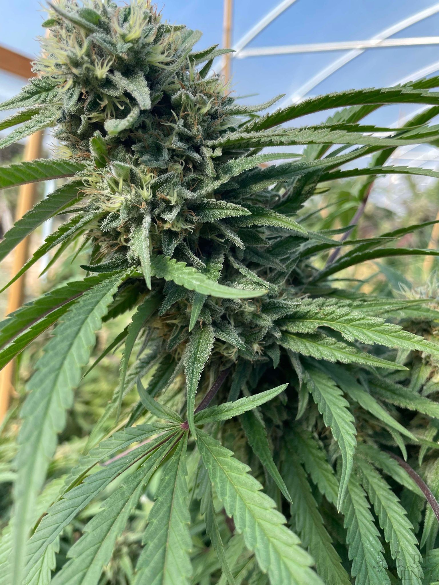
– Cannabis (hemp)
– Humulus (hops)
– Celtis (hackberries)
Celtis is the largest genus, containing about 100 species[1].
Characteristics
Members of the Cannabaceae family exhibit diverse characteristics:
– Growth forms: Can be trees, erect herbs, or twining herbs[1]
– Leaves: Often palmately lobed or compound, with stipules[1]
– Flowers: Typically small, wind-pollinated, and lacking petals[1][2]
– Fruits: Usually dry, one-seeded achenes or drupes[2]
Many Cannabaceae species are dioecious, meaning they have separate male and female plants[1].
Economic Importance
The most economically significant genera in Cannabaceae are:
1. Cannabis: Used for hemp fiber, seeds, oils, medicinal purposes, and as a recreational drug[4]
2. Humulus: Primarily used in beer production[4]
3. Celtis: Valued for timber and ornamental purposes[2]
Evolutionary History
Cannabaceae likely originated in East Asia during the Late Cretaceous period, approximately 94-90 million years ago[1][3]. Fossils indicate that the family was widely distributed in the Northern Hemisphere during the early Cenozoic era, later shifting towards more tropical regions due to climate changes[1][3].
Interesting Facts
– Cannabis and hops (Humulus) are genetically related, both belonging to the Cannabaceae family[4]
– The family was previously classified under the order Urticales but is now placed within Rosales based on molecular data[1]
– Some Cannabaceae species have been used in traditional medicine[4]
Citations:
[1] https://en.wikipedia.org/wiki/Cannabaceae
[2] https://www.britannica.com/plant/Cannabaceae
[3] https://www.gbif.org/species/165599050
[4] https://herbliz.de/en/blog/cbd-dictionary/cannabaceae/
[5] https://en.wikipedia.org/wiki/Cannabis
[6] https://florida.plantatlas.usf.edu/Family.aspx?id=57
[7] https://www.sciencedirect.com/topics/agricultural-and-biological-sciences/cannabaceae
[8] https://www.sciencedirect.com/topics/biochemistry-genetics-and-molecular-biology/cannabaceae
Well, that’s interesting to know that Psilotum nudum are known as whisk ferns. Psilotum nudum is the commoner species of the two. While the P. flaccidum is a rare species and is found in the tropical islands. Both the species are usually epiphytic in habit and grow upon tree ferns. These species may also be terrestrial and grow in humus or in the crevices of the rocks.
View the detailed Guide of Psilotum nudum: Detailed Study Of Psilotum Nudum (Whisk Fern), Classification, Anatomy, Reproduction