
Size of this PNG preview of this SVG file: 800 × 514 pixels. Other resolutions: 320 × 206 pixels | 640 × 411 pixels | 1,024 × 658 pixels | 1,280 × 823 pixels | 2,560 × 1,646 pixels | 1,047 × 673 pixels.
Original file (SVG file, nominally 1,047 × 673 pixels, file size: 291 KB)
Summary
| Description |
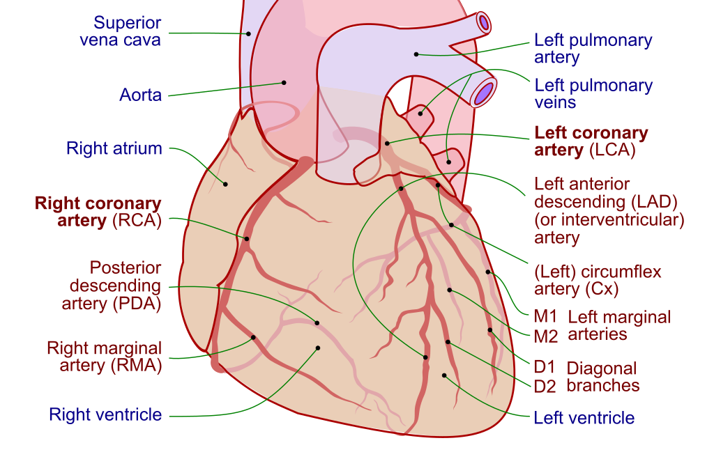
English: Coronary circulation, with coronary arteries labeled in red text and other landmarks in blue text.
This vector graphics image was originally created with Adobe Illustrator, and modified with Inkscape. |
||
| Date | |||
| Source | |||
| Author |
|
||
| Permission (Reusing this file) |
This file is licensed under the Creative Commons Attribution-Share Alike 3.0 Unported license.
|
||
| Other versions |
[] SVG
PNG
|
||
| SVG development | This diagram was created with Inkscape. This diagram uses embedded text that can be easily translated using a text editor.
|
.jpg)


Original upload log
This image is a derivative work of the following images:
- Coronary.pdf licensed with Cc-by-sa-3.0,2.5,2.0,1.0, GFDL
- 2007-10-05T21:05:19Z Toony 1727x2011 (129568 Bytes) {{Information |Description=Coronary |Source=[[Image:Heart left anterior oblique diagrams.svg|100px]] |Date=2007 |Author=Patrick J. Lynch, medical illustrator |Permission= |other_versions= }}
Uploaded with derivativeFX
File history
Click on a date/time to view the file as it appeared at that time.
| Date/Time | Thumbnail | Dimensions | User | Comment | |
|---|---|---|---|---|---|
| current | 12:48, 28 June 2021 | | 1,047 × 673 (291 KB) | Jmarchn | Validation W3C |
| 17:33, 23 November 2020 |  | 1,047 × 673 (366 KB) | Jmarchn | Fixed minor arrow error | |
| 22:59, 22 November 2020 |  | 1,047 × 673 (366 KB) | Jmarchn | Solved pulmonary artery line too thin | |
| 22:55, 22 November 2020 |  | 1,047 × 673 (366 KB) | Jmarchn | Better draw: Added Pulmonary artery, blue color for venous blood and text to path layer. | |
| 17:16, 22 November 2020 |  | 1,047 × 673 (95 KB) | Jmarchn | Solved label alignment problem | |
| 17:05, 22 November 2020 |  | 1,047 × 673 (97 KB) | Jmarchn | Better draw. According https://radiologyassistant.nl/cardiovascular/anatomy/coronary-anatomy-and-anomalies and more. | |
| 07:19, 4 September 2010 |  | 512 × 294 (79 KB) | Mikael Häggström | Correction of marginal arteries | |
| 06:39, 4 September 2010 |  | 512 × 294 (79 KB) | Mikael Häggström | Small adaption to avoid text getting out of frame | |
| 06:27, 4 September 2010 |  | 512 × 298 (79 KB) | Mikael Häggström | Adapted in Inkscape to be more consistent with other images of coronary arteries (Human heart with coronary arteries new.png, [http://www.jonbarron.org/content/images/CoronariesFrontal. | |
| 15:55, 9 April 2010 |  | 512 × 563 (48 KB) | Fred the Oyster | {{Information |Description=Basic illustration of positioning of aorta, pulmonary vein and coronary arteries |Source=*File:Coronary.pdf |Date=2010-04-09 15:55 (UTC) |Author=*File:Coronary.pdf: Patrick J. Lynch, medical illustrator *derivative w |
File usage
The following pages on the English Wikipedia use this file (pages on other projects are not listed):
- Cardiac allograft vasculopathy
- Circumflex branch of left coronary artery
- Coronary arteries
- Coronary artery aneurysm
- Coronary circulation
- Coronary ischemia
- Drug-eluting stent
- Heart
- Left anterior descending artery
- Left coronary artery
- Left marginal artery
- Percutaneous coronary intervention
- Posterior descending artery
- Right coronary artery
- Right marginal branch of right coronary artery
Global file usage
The following other wikis use this file:
- Usage on ar.wikipedia.org
- Usage on cs.wikipedia.org
- Usage on de.wikipedia.org
- Usage on el.wikipedia.org
- Usage on fa.wikipedia.org
- Usage on fi.wikipedia.org
- Usage on hi.wikipedia.org
- Usage on hr.wikipedia.org
- Usage on hy.wikipedia.org
- Usage on id.wikipedia.org
- Usage on ja.wikipedia.org
- Usage on ko.wikipedia.org
- Usage on lv.wikipedia.org
- Usage on mk.wikipedia.org
- Usage on ms.wikipedia.org
- Usage on new.wikipedia.org
- Usage on nl.wikipedia.org
- Usage on ro.wikipedia.org
- Usage on si.wikipedia.org
- Usage on sk.wikipedia.org
- Usage on sv.wikipedia.org
- Usage on te.wikipedia.org
- Usage on th.wikipedia.org
- Usage on tr.wikipedia.org
- Usage on uk.wikipedia.org
- Usage on zh.wikipedia.org