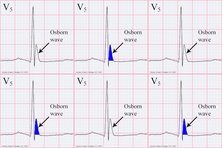

A J wave — also known as Osborn wave, camel-hump sign, late delta wave, hathook junction, hypothermic wave,[1] K wave, H wave or current of injury — is an abnormal electrocardiogram finding.[2]
J waves are positive deflections occurring at the junction between the QRS complex and the ST segment,[3][4] where the S point, also known as the J point, has a myocardial infarction-like elevation.
Causes[edit]
They are usually observed in people suffering from hypothermia with a temperature of less than 32 °C (90 °F),[5] though they may also occur in people with very high blood levels of calcium (hypercalcemia), brain injury, vasospastic angina, acute pericarditis, or they could also be a normal variant.[citation needed] Osborn waves on ECG are frequent during targeted temperature management (TTM) after cardiac arrest, particularly in patients treated with 33 °C.[6] Osborn waves are not associated with increased risk of ventricular arrhythmia, and may be considered a benign physiological phenomenon, associated with lower mortality in univariable analyses.[6]
History[edit]
The prominent J deflection attributed to hypothermia was first reported in 1938 by Tomaszewski. These waves were then definitively described in 1953 by John J. Osborn (1917–2014) and were named in his honor.[7] Over time, the wave has increasingly been referred to as a J wave, though is still sometimes referred to as the Osborn wave in most part due to Osborn's article in the American Journal of Physiology on experimental hypothermia.[8]
References[edit]
- ^ Aydin M, Gursurer M, Bayraktaroglu T, Kulah E, Onuk T (2005). "Prominent J wave (Osborn wave) with coincidental hypothermia in a 64-year-old woman". Tex Heart Inst J. 32 (1): 105. PMC 555838. PMID 15902836.
- ^ Maruyama M, Kobayashi Y, Kodani E, et al. (2004). "Osborn waves: history and significance". Indian Pacing Electrophysiol J. 4 (1): 33–9. PMC 1501063. PMID 16943886.
- ^ "ecg_6lead018.html". Retrieved 2008-12-20.
- ^ "THE MERCK MANUAL OF GERIATRICS, Ch. 67, Hyperthermia and Hypothermia, Fig. 67-1". Retrieved 2008-12-20.
- ^ Marx, John (2010). Rosen's emergency medicine: concepts and clinical practice 7th edition. Philadelphia, PA: Mosby/Elsevier. p. 1869. ISBN 978-0-323-05472-0.
- ^ a b Hadziselimovic, Edina; Thomsen, Jakob Hartvig; Kjaergaard, Jesper; Køber, Lars; Graff, Claus; Pehrson, Steen; Nielsen, Niklas; Erlinge, David; Frydland, Martin; Wiberg, Sebastian; Hassager, Christian (July 2018). "Osborn waves following out-of-hospital cardiac arrest—Effect of level of temperature management and risk of arrhythmia and death". Resuscitation. 128: 119–125. doi:10.1016/j.resuscitation.2018.04.037. PMID 29723608.
- ^ Osborn, J. J. (1953). "Experimental hypothermia: Respiratory and blood pH changes in relation to cardiac function". Am J Physiol. 175 (3): 389–398. doi:10.1152/ajplegacy.1953.175.3.389. PMID 13114420.
- ^ Serafi, S.; Vliek, C.; Taremi, M. (2011). "Osborn waves in a hypothermic patient". Journal of Community Hospital Internal Medicine Perspectives. 1 (4). Article: 10742. doi:10.3402/jchimp.v1i4.10742. PMC 3714046. PMID 23882340.
Well, that’s interesting to know that Psilotum nudum are known as whisk ferns. Psilotum nudum is the commoner species of the two. While the P. flaccidum is a rare species and is found in the tropical islands. Both the species are usually epiphytic in habit and grow upon tree ferns. These species may also be terrestrial and grow in humus or in the crevices of the rocks.
View the detailed Guide of Psilotum nudum: Detailed Study Of Psilotum Nudum (Whisk Fern), Classification, Anatomy, Reproduction