| Parieto-occipital sulcus | |
|---|---|
 Fig. 726: Lateral surface of left cerebral hemisphere, viewed from the side. | |
 Fig. 727: Medial surface of left cerebral hemisphere. | |
| Details | |
| Identifiers | |
| Latin | sulcus parietooccipitalis, fissura parietooccipitalis |
| NeuroNames | 52 |
| NeuroLex ID | birnlex_1428 |
| TA98 | A14.1.09.108 |
| TA2 | 5437 |
| FMA | 83754 |
| Anatomical terms of neuroanatomy | |
In neuroanatomy, the parieto-occipital sulcus (also called the parieto-occipital fissure) is a deep sulcus in the cerebral cortex that marks the boundary between the cuneus and precuneus, and also between the parietal and occipital lobes. Only a small part can be seen on the lateral surface of the hemisphere, its chief part being on the medial surface.
The lateral part of the parieto-occipital sulcus (Fig. 726) is situated about 5 cm in front of the occipital pole of the hemisphere, and measures about 1.25 cm. in length.
The medial part of the parieto-occipital sulcus (Fig. 727) runs downward and forward as a deep cleft on the medial surface of the hemisphere, and joins the calcarine fissure below and behind the posterior end of the corpus callosum. In most cases, it contains a submerged gyrus.
Function[edit]
The parieto-occipital lobe has been found in various neuroimaging studies, including PET (positron-emission-tomography) studies,[1][2][3][4] and SPECT (single-photon emission computed tomography) studies,[5][6] to be involved along with the dorsolateral prefrontal cortex during planning.
Gallery[edit]
-
 Animation of left cerebral hemisphere. Parieto-occipital sulcus shown in red.
 -
 Medial surface of right hemisphere. Parieto-occipital sulcus labeled at top right as "*"
 -
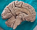 Medial surface of left hemisphere. Parieto-occipital sulcus visible at top left.
 -
Human brain dissection video (1 min 52 sec). Demonstrating location of parieto-occipital sulcus of left cerebral hemisphere.


References[edit]
![]() This article incorporates text in the public domain from page 820 of the 20th edition of Gray's Anatomy (1918)
This article incorporates text in the public domain from page 820 of the 20th edition of Gray's Anatomy (1918)
- ^ Owen, Adrian M.; Doyon, Julien; Petrides, Michael; Evans, Alan C. (1996). "Planning and Spatial Working Memory: a Positron Emission Tomography Study in Humans". European Journal of Neuroscience. 8 (2): 353–364. doi:10.1111/j.1460-9568.1996.tb01219.x. PMID 8714706. S2CID 21770063.
- ^ Baker, S.C.; Rogers, R.D.; Owen, A.M.; Frith, C.D.; Dolan, R.J.; Frackowiak, R.S.J.; Robbins, T.W. (June 1996). "Neural Systems Engaged by Planning: a PET Study of the Tower of London Task" (PDF). Neuropsychologia. 34 (6): 515–526. doi:10.1016/0028-3932(95)00133-6. hdl:21.11116/0000-0001-A39D-6. PMID 8736565. S2CID 7366716. Retrieved 18 June 2014.
- ^ Dagher, Alain; Owen, Adrian M.; Boecker, Henning; Brooks, David J. (October 1999). "Mapping the Network for Planning: a Correlational PET activation study with the Tower of London Task". Brain. 122 (10): 1973–1987. doi:10.1093/brain/122.10.1973. PMID 10506098.
- ^ Rowe, J.B.; Owen, Adrian M.; Johnsrude, Ingrid S.; Passingham, R.E. (2001). "Imaging the Mental Components of a Planning Task" (PDF). Neuropsychologia. 39 (3): 315–327. doi:10.1016/S0028-3932(00)00109-3. PMID 11163609. S2CID 21819578. Retrieved 18 June 2014.
- ^ Rezai, Karim; Andreasen, Nancy C.; Alliger, Randy; Cohen, Gregg; Swayze, Victor II; O'Leary, Daniel S. (June 1993). "The Neuropsychology of the Prefrontal Cortex". Archives of Neurology. 50 (6): 636–642. doi:10.1001/archneur.1993.00540060066020. PMID 8503801. Retrieved 18 June 2014.
- ^ Morris, R.G.; Ahmed, S.; Syed, G.M.; Toone, B.K. (December 1993). "Neural Correlates of Planning Ability: Frontal Lobe Activation during the Tower of London Test". Neuropsychologia. 31 (12): 1367–1378. doi:10.1016/0028-3932(93)90104-8. PMID 8127433. S2CID 13351226.
External links[edit]
- "Anatomy diagram: 13048.000-3". Roche Lexicon - illustrated navigator. Elsevier. Archived from the original on 2012-07-22.
- https://web.archive.org/web/20070316043257/http://www2.umdnj.edu/~neuro/studyaid/Practical2000/Q30.htm