| Acinar cell carcinoma of the pancreas | |
|---|---|
| Other names | Acinar cell carcinoma |
 | |
| Micrograph of an acinar cell carcinoma of the pancreas. H&E stain. | |
| Specialty | Oncology |
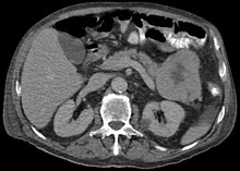
Acinar cell carcinoma of the pancreas, also acinar cell carcinoma, is a rare malignant exocrine tumour of the pancreas. It represents 5% of all exocrine tumours of the pancreas, making it the second most common type of pancreatic cancer.[1] It is abbreviated ACC. It typically has a guarded prognosis.
Signs and symptoms[edit]

The disease is more common in men than women and the average age at diagnosis is about 60.[2] Symptoms are often non-specific and include weight loss. A classic presentation, found in around 15% of cases includes subcutaneous nodules (due to fat necrosis) and arthralgias, caused by a release of lipase.[2]
Pathology[edit]
ACC is associated with increased serum lipase and manifests in the classic presentation known as the Schmid triad (subcutaneous fat necrosis, polyarthritis, eosinophilia).[3]
ACC are typically large, up to 10 cm, and soft compared to pancreatic adenocarcinoma, lacking its dense stroma. They can arise in any part of the pancreas.[2]
Histomorphologically, the tumour resembles the cells of the pancreatic acini and, typically, have moderate granular cytoplasm that stain with both PAS and PASD.[4]
Diagnosis[edit]


Light microscopy of an acinar cell carcinoma biopsy typically shows granular appearance.[6] Immunohistochemistry is usually positive for trypsin, chymotrypsin and lipase.[6] On genetic testing, altered genes/proteins are typically found for p53, SMAD4, APC, ARID1A and GNAS.[6]
Treatment[edit]
ACC can be treated with a Whipple procedure or (depending on the location within the pancreas) with left partial resection of pancreas.[citation needed]
See also[edit]
References[edit]
- ^ Tobias Jeffrey S., Hochhauser, Daniel, Cancer and its Management, p. 276, 2010 (6th edn), ISBN 1118713257, 9781118713259
- ^ a b c Von Hoff": Daniel D. Von Hoff, Douglas Brian Evans, Ralph H. Hruban, eds. Pancreatic Cancer, 2005, Jones & Bartlett Learning, ISBN 0763721786, 9780763721787
- ^ Jang, SH.; Choi, SY.; Min, JH.; Kim, TW.; Lee, JA.; Byun, SJ.; Lee, JW. (Feb 2010). "[A case of acinar cell carcinoma of pancreas, manifested by subcutaneous nodule as initial clinical symptom]". Korean J Gastroenterol. 55 (2): 139–43. doi:10.4166/kjg.2010.55.2.139. PMID 20168061.
- ^ Klimstra, DS.; Heffess, CS.; Oertel, JE.; Rosai, J. (Sep 1992). "Acinar cell carcinoma of the pancreas. A clinicopathologic study of 28 cases". Am J Surg Pathol. 16 (9): 815–37. doi:10.1097/00000478-199209000-00001. PMID 1384374. S2CID 19317244.
- ^ Wang Y, Miller FH, Chen ZE, Merrick L, Mortele KJ, Hoff FL; et al. (2011). "Diffusion-weighted MR imaging of solid and cystic lesions of the pancreas". Radiographics. 31 (3): E47-64. doi:10.1148/rg.313105174. PMID 21721197.
{{cite journal}}: CS1 maint: multiple names: authors list (link)
Diagram by Mikael Häggström, M.D. - ^ a b c Pishvaian MJ, Brody JR (2017). "Therapeutic Implications of Molecular Subtyping for Pancreatic Cancer". Oncology (Williston Park). 31 (3): 159–66, 168. PMID 28299752.