| LIPC | |||||||||||||||||||||||||||||||||||||||||||||||||||
|---|---|---|---|---|---|---|---|---|---|---|---|---|---|---|---|---|---|---|---|---|---|---|---|---|---|---|---|---|---|---|---|---|---|---|---|---|---|---|---|---|---|---|---|---|---|---|---|---|---|---|---|
 | |||||||||||||||||||||||||||||||||||||||||||||||||||
| Identifiers | |||||||||||||||||||||||||||||||||||||||||||||||||||
| Aliases | LIPC, HDLCQ12, HL, HTGL, LIPH, lipase C, hepatic type | ||||||||||||||||||||||||||||||||||||||||||||||||||
| External IDs | OMIM: 151670 MGI: 96216 HomoloGene: 199 GeneCards: LIPC | ||||||||||||||||||||||||||||||||||||||||||||||||||
| |||||||||||||||||||||||||||||||||||||||||||||||||||
| |||||||||||||||||||||||||||||||||||||||||||||||||||
| |||||||||||||||||||||||||||||||||||||||||||||||||||
| |||||||||||||||||||||||||||||||||||||||||||||||||||
| |||||||||||||||||||||||||||||||||||||||||||||||||||
| Wikidata | |||||||||||||||||||||||||||||||||||||||||||||||||||
| |||||||||||||||||||||||||||||||||||||||||||||||||||
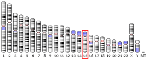
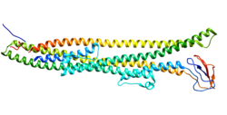
Hepatic lipase (HL), also called hepatic triglyceride lipase (HTGL) or LIPC (for "lipase, hepatic"), is a form of lipase, catalyzing the hydrolysis of triacylglyceride. Hepatic lipase is coded by chromosome 15 and its gene is also often referred to as HTGL or LIPC.[5] Hepatic lipase is expressed mainly in liver cells, known as hepatocytes, and endothelial cells of the liver. The hepatic lipase can either remain attached to the liver or can unbind from the liver endothelial cells and is free to enter the body's circulation system.[6] When bound on the endothelial cells of the liver, it is often found bound to heparan sulfate proteoglycans (HSPG), keeping HL inactive and unable to bind to HDL (high-density lipoprotein) or IDL (intermediate-density lipoprotein).[7] When it is free in the bloodstream, however, it is found associated with HDL to maintain it inactive. This is because the triacylglycerides in HDL serve as a substrate, but the lipoprotein contains proteins around the triacylglycerides that can prevent the triacylglycerides from being broken down by HL.[8]
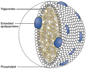
One of the principal functions of hepatic lipase is to convert intermediate-density lipoprotein (IDL) to low-density lipoprotein (LDL). Hepatic lipase thus plays an important role in triglyceride level regulation in the blood by maintaining steady levels of IDL, HDL and LDL.[5]

Function[edit]
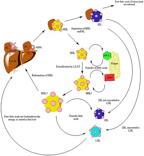
Hepatic lipase falls under a class of enzymes known as hydrolases. Its function is to hydrolyze triacylglycerol to diacylglycerol and carboxylate (free fatty acids) with the addition of water.[10] The substrate, triacylglycerol, comes from IDL (intermediate-density lipoprotein) and the release of free fatty acids converts IDL into LDL (low-density lipoprotein).[7] These remaining remnants of LDL can be sent back to the liver, where it can be stored for later use or broken down to harness its energy. It can also be sent to peripheral cells for its cholesterol and used in anabolic pathways to build molecules that the cell needs such as hormones that include a cholesterol backbone.[11]
To prevent the build-up of plaque (also referred to as a lipid pool), nascent HDL molecules which are low in triglycerides, take off free fatty acids from the plaques through the help of ABCL1 proteins. These proteins help transfer free fatty acids from plaques in the arteries to HDL.[8] This process creates HDL3 (High density lipoprotein 3), a mature HDL molecule that has been esterified by another enzyme known as LCAT.[11] More free fatty acids can be taken up from the plaque by SR-B1 receptors, which convert HDL3 to HDL2 which contains higher concentrations of free fatty acids.[7] HDL2 can then interact with LDL and IDL by transferring over fatty acids that have accumulated in the plaque. Hepatic lipase can then catalyze the conversion of IDL to LDL by breaking down triacylglycerides in IDL and release free fatty acids to be used by other cells with low concentrations of cholesterol or stored in the liver for later use.[8]
Hepatic lipase regulation[edit]
The human body contains two inactive forms of HL. One inactive form is found on the liver bound to HSPG (heparin sulfate proteoglycans) and the second inactive form is found in the blood bound to HDL, inactivated by the proteins on the surface of the lipoprotein. The activation of HL occurs in two steps. First, HDL that makes its way to the liver, binds to HL thereby removing the heparan sulfate proteoglycan and freeing up the hepatic lipase into the bloodstream, but HL is still inactive due to the proteins on the surface of the lipoprotein. Second, HDL unbinds from HL to activate HL enzymes in the blood.[6]
HDL has been found to be regulated by electrostatic interactions with lipoproteins such as HDL. When HDL takes up free fatty acids from cells to prevent plaque build-up, it begins to increase its overall negative charge and instead stimulates HL to catalysis the triacylglycerides inside of VLDL (very low density lipoprotein). This is because the build-up of negative charge in HDL inhibits binding, but will allow HL to catalyze other lipoproteins. Other lipoproteins, such as ApoE, works in a similar way by inhibiting binding of HL and HDL but will allow HL to catalyze other lipoproteins.[8]
Other factors that contribute the regulation of HL are due to sex differences between women and men. It has been shown that women contain lower levels of ApoE along with an increased amount of free HL enzymes in their circulatory system when compared to men. The production of estrogen in women is also believed to reduce the activity of HL by serving as an inhibitor of gene transcription.[7]
Secretion of HDL from the liver into the circulatory system regulates the release of HL into the body's bloodstream. This is because factors that increase the release of HDL (such as fasting, leading to low levels of HDL) increases the amount of HL bound to HDL and released into the bloodstream. Another lipoprotein, ApoA-I, which increases release of HDL was shown to have a similar effect by mutating the gene that coded it. Mutated ApoA-I protein caused a decrease in HL release and thus decreased the amount of HL bound to HDL and released into the bloodstream.[7]
Clinical significance[edit]
Hepatic lipase deficiency is a rare, autosomal recessive disorder that results in elevated high-density lipoprotein (HDL) cholesterol due to a mutation in the hepatic lipase gene. Clinical features are not well understood and there are no characteristic xanthomas. There is an association with a delay in atherosclerosis in an animal model.[6]
In many studies that have been conducted, hepatic lipase is also closely related to obesity. In one test, an experiment was created by Cedó et al. where mouse cells were created to have a mutated HL protein that has lost its function. They found that a build-up of triglyceride levels led to nonalcoholic fatty liver disease. This was due to HL's inability to convert the triacylglycerides in IDL, and thereby creating LDL. Thus, the inability of endothelial cells to take up free fatty acids becomes higher and more IDL gets stored in the liver. This deficiency in HL also leads to liver inflammation and obesity problems. In the experiment though, mouse HL is found unbound to heparan sulfate proteoglycans (HSPG) whereas human HL is found attached to heparan sulfate proteoglycans (HSPG), deactivating HL until bound to IDL. More experiments must be performed to determine the potential effects in humans.[8]
References[edit]
- ^ a b c GRCh38: Ensembl release 89: ENSG00000166035 - Ensembl, May 2017
- ^ a b c GRCm38: Ensembl release 89: ENSMUSG00000032207 - Ensembl, May 2017
- ^ "Human PubMed Reference:". National Center for Biotechnology Information, U.S. National Library of Medicine.
- ^ "Mouse PubMed Reference:". National Center for Biotechnology Information, U.S. National Library of Medicine.
- ^ a b Fox SI (2015). Human physiology (Fourteenth ed.). New York, NY. ISBN 978-0-07-783637-5. OCLC 895500922.
{{cite book}}: CS1 maint: location missing publisher (link) - ^ a b c Karackattu SL, Trigatti B, Krieger M (March 2006). "Hepatic lipase deficiency delays atherosclerosis, myocardial infarction, and cardiac dysfunction and extends lifespan in SR-BI/apolipoprotein E double knockout mice". Arteriosclerosis, Thrombosis, and Vascular Biology. 26 (3): 548–54. doi:10.1161/01.ATV.0000202662.63876.02. PMID 16397139. S2CID 336426.
- ^ a b c d e Chatterjee C, Sparks DL (April 2011). "Hepatic lipase, high density lipoproteins, and hypertriglyceridemia". The American Journal of Pathology. 178 (4): 1429–33. doi:10.1016/j.ajpath.2010.12.050. PMC 3078429. PMID 21406176.
- ^ a b c d e Cedó L, Santos D, Roglans N, Julve J, Pallarès V, Rivas-Urbina A, Llorente-Cortes V, Laguna JC, Blanco-Vaca F, Escolà-Gil JC (2017). "Human hepatic lipase overexpression in mice induces hepatic steatosis and obesity through promoting hepatic lipogenesis and white adipose tissue lipolysis and fatty acid uptake". PLOS ONE. 12 (12): e0189834. Bibcode:2017PLoSO..1289834C. doi:10.1371/journal.pone.0189834. PMC 5731695. PMID 29244870.
- ^ Bourne Y, Cambillau C (1994). "Horse Pancreatic Lipase. The Crystal Structure at 2.3 Angstroms Resolution". J. Mol. Biol. 238. doi:10.2210/pdb1hpl/pdb.
- ^ "Hepatic triacylglycerol lipase - P11150 (LIPC_HUMAN)". RCSB Protein Data Bank. Archived from the original on 2018-06-12. Retrieved 2018-02-07.
- ^ a b Sanan DA, Fan J, Bensadoun A, Taylor JM (May 1997). "Hepatic lipase is abundant on both hepatocyte and endothelial cell surfaces in the liver". Journal of Lipid Research. 38 (5): 1002–13. doi:10.1016/S0022-2275(20)37224-2. PMID 9186917.
Further reading[edit]
- Santamarina-Fojo S, Haudenschild C, Amar M (June 1998). "The role of hepatic lipase in lipoprotein metabolism and atherosclerosis". Current Opinion in Lipidology. 9 (3): 211–9. doi:10.1097/00041433-199806000-00005. PMID 9645503.
- Jansen H, Verhoeven AJ, Sijbrands EJ (September 2002). "Hepatic lipase: a pro- or anti-atherogenic protein?". Journal of Lipid Research. 43 (9): 1352–62. doi:10.1194/jlr.R200008-JLR200. hdl:1765/9973. PMID 12235167.
- Zambon A, Deeb SS, Pauletto P, Crepaldi G, Brunzell JD (April 2003). "Hepatic lipase: a marker for cardiovascular disease risk and response to therapy". Current Opinion in Lipidology. 14 (2): 179–89. doi:10.1097/00041433-200304000-00010. PMID 12642787. S2CID 23377060.
- Hegele RA, Tu L, Connelly PW (1993). "Human hepatic lipase mutations and polymorphisms". Human Mutation. 1 (4): 320–4. doi:10.1002/humu.1380010410. PMID 1301939. S2CID 45428213.
- Hegele RA, Vezina C, Moorjani S, Lupien PJ, Gagne C, Brun LD, Little JA, Connelly PW (March 1991). "A hepatic lipase gene mutation associated with heritable lipolytic deficiency". The Journal of Clinical Endocrinology and Metabolism. 72 (3): 730–2. doi:10.1210/jcem-72-3-730. PMID 1671786.
- Hegele RA, Little JA, Connelly PW (August 1991). "Compound heterozygosity for mutant hepatic lipase in familial hepatic lipase deficiency". Biochemical and Biophysical Research Communications. 179 (1): 78–84. doi:10.1016/0006-291X(91)91336-B. PMID 1883393.
- Ameis D, Stahnke G, Kobayashi J, McLean J, Lee G, Büscher M, Schotz MC, Will H (April 1990). "Isolation and characterization of the human hepatic lipase gene". The Journal of Biological Chemistry. 265 (12): 6552–5. doi:10.1016/S0021-9258(19)39182-3. PMID 2324091.
- Datta S, Luo CC, Li WH, VanTuinen P, Ledbetter DH, Brown MA, Chen SH, Liu SW, Chan L (January 1988). "Human hepatic lipase. Cloned cDNA sequence, restriction fragment length polymorphisms, chromosomal localization, and evolutionary relationships with lipoprotein lipase and pancreatic lipase". The Journal of Biological Chemistry. 263 (3): 1107–10. doi:10.1016/S0021-9258(19)57271-4. PMID 2447084.
- Cai SJ, Wong DM, Chen SH, Chan L (November 1989). "Structure of the human hepatic triglyceride lipase gene". Biochemistry. 28 (23): 8966–71. doi:10.1021/bi00449a002. PMID 2605236.
- Stahnke G, Sprengel R, Augustin J, Will H (1988). "Human hepatic triglyceride lipase: cDNA cloning, amino acid sequence and expression in a cultured cell line". Differentiation; Research in Biological Diversity. 35 (1): 45–52. doi:10.1111/j.1432-0436.1987.tb00150.x. PMID 2828141.
- Martin GA, Busch SJ, Meredith GD, Cardin AD, Blankenship DT, Mao SJ, Rechtin AE, Woods CW, Racke MM, Schafer MP (August 1988). "Isolation and cDNA sequence of human postheparin plasma hepatic triglyceride lipase". The Journal of Biological Chemistry. 263 (22): 10907–14. doi:10.1016/S0021-9258(18)38056-6. PMID 2839510.
- Sparkes RS, Zollman S, Klisak I, Kirchgessner TG, Komaromy MC, Mohandas T, Schotz MC, Lusis AJ (October 1987). "Human genes involved in lipolysis of plasma lipoproteins: mapping of loci for lipoprotein lipase to 8p22 and hepatic lipase to 15q21". Genomics. 1 (2): 138–44. doi:10.1016/0888-7543(87)90005-X. PMID 3692485.
- Kounnas MZ, Chappell DA, Wong H, Argraves WS, Strickland DK (April 1995). "The cellular internalization and degradation of hepatic lipase is mediated by low density lipoprotein receptor-related protein and requires cell surface proteoglycans". The Journal of Biological Chemistry. 270 (16): 9307–12. doi:10.1074/jbc.270.16.9543. PMID 7721852.
- Mori A, Takagi A, Ikeda Y, Ashida Y, Yamamoto A (August 1996). "An AvaII polymorphism in exon 5 of the human hepatic triglyceride lipase gene". Molecular and Cellular Probes. 10 (4): 309–11. doi:10.1006/mcpr.1996.0040. PMID 8865179.
- Takagi A, Ikeda Y, Mori A, Ashida Y, Yamamoto A (August 1996). "Identification of a BstNI polymorphism in exon 9 of the human hepatic triglyceride lipase gene". Molecular and Cellular Probes. 10 (4): 313–4. doi:10.1006/mcpr.1996.0041. PMID 8865180.
- Choi SY, Goldberg IJ, Curtiss LK, Cooper AD (August 1998). "Interaction between ApoB and hepatic lipase mediates the uptake of ApoB-containing lipoproteins". The Journal of Biological Chemistry. 273 (32): 20456–62. doi:10.1074/jbc.273.32.20456. PMID 9685400.
- Cargill M, Altshuler D, Ireland J, Sklar P, Ardlie K, Patil N, Shaw N, Lane CR, Lim EP, Kalyanaraman N, Nemesh J, Ziaugra L, Friedland L, Rolfe A, Warrington J, Lipshutz R, Daley GQ, Lander ES (July 1999). "Characterization of single-nucleotide polymorphisms in coding regions of human genes". Nature Genetics. 22 (3): 231–8. doi:10.1038/10290. PMID 10391209. S2CID 195213008.
- Tiebel O, Gehrisch S, Pietzsch J, Gromeier S, Jaross W (2000). "18 bp insertion/duplication with internal missense mutation in human hepatic lipase gene exon 3. Mutations in brief no. 181. Online". Human Mutation. 12 (3): 216. PMID 10660332.
- Yamakawa-Kobayashi K, Somekawa Y, Fujimura M, Tomura S, Arinami T, Hamaguchi H (May 2002). "Relation of the -514C/T polymorphism in the hepatic lipase gene to serum HDL and LDL cholesterol levels in postmenopausal women under hormone replacement therapy". Atherosclerosis. 162 (1): 17–21. doi:10.1016/S0021-9150(01)00675-X. PMID 11947893.
External links[edit]
- hepatic+lipase,+human at the U.S. National Library of Medicine Medical Subject Headings (MeSH)
