Trichome

Original file(1,595 × 1,594 pixels, file size: 776 KB, MIME type: image/jpeg)

Summary

Description
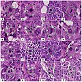
English: Snapshots from several WSI of several breast resections diagnosed previously as an infiltrative ductal carcinoma using a light microscopy. These snapshots showing different appearances of mitotic figures encircled by green circles. Panels A–C show cells in early metaphase. Panels D–G show different forms of mitotic division in late metaphase. Panel H–L shows different forms of anaphase. Panels M–P show cells in telophase.
Date
Source

(2013). "Evaluation of Mitotic Activity Index in Breast Cancer Using Whole Slide Digital Images". PLoS ONE 8 (12): e82576. DOI:10.1371/journal.pone.0082576. ISSN 1932-6203.

Author Al-Janabi S, van Slooten H-J, Visser M, van der Ploeg T, van Diest PJ, Jiwa M

Licensing

w:en:Creative Commons
attribution
This file is licensed under the Creative Commons Attribution 4.0 International license.
You are free:
  • to share – to copy, distribute and transmit the work
  • to remix – to adapt the work
Under the following conditions:
  • attribution – You must give appropriate credit, provide a link to the license, and indicate if changes were made. You may do so in any reasonable manner, but not in any way that suggests the licensor endorses you or your use.

Captions

Mitosis appearances in breast cancer

Items portrayed in this file

depicts

copyright status

copyrighted

copyright license

Creative Commons Attribution 4.0 International

inception

30 December 2013

File history

Click on a date/time to view the file as it appeared at that time.

Date/TimeThumbnailDimensionsUserComment
current11:45, 4 October 2019Thumbnail for version as of 11:45, 4 October 20191,595 × 1,594 (776 KB)Mikael HäggströmUser created page with UploadWizard
The following pages on the English Wikipedia use this file (pages on other projects are not listed):

Global file usage

The following other wikis use this file:

Leave a Reply