Size of this preview: 398 × 600 pixels. Other resolutions: 159 × 240 pixels | 446 × 672 pixels.
Original file (446 × 672 pixels, file size: 1.65 MB, MIME type: image/gif, looped, 32 frames, 6.4 s)
Summary
| Description |
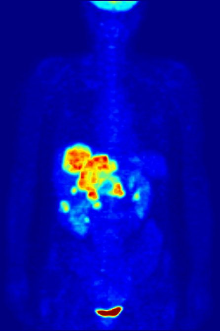
English: Maximum Intensity Projection (MIP) of a wholebody positron emission tomography (PET) acquisition of a 79 kg (174 lb) weighting female after intravenous injection of 371 MBq of 18F-FDG (one hour prior measurement). The investigation has been performed as part of a tumor diagnosis prior to applying a radiotherapy (tumor staging step). Besides normal accumulation of the tracer in the heart, bladder, kidneys and brain, liver metastases of a colorectal tumor are clearly visible within the abdominal region of the image.
Deutsch: Maximumintensitätsprojektion (MIP) einer Ganzkörperaufnahme mittels Positronen-Emissions-Tomographie (PET). Die Aufnahme zeigt eine 79 kg schwere weibliche Patientin nach intravenöser Injektion von 371 MBq 18F-FDG (eine Stunde vor Messung). Die Untersuchung wurde im Rahmen einer Tumordiagnose vor Anwendung einer Strahlentherapie (sogn. Tumorstaging) d88urchgeführt. Neben den normalen Anreicherungen des Tracers in Herz, Blase, Nieren und Gehirn, sind auch Lebermetastasen eines kolorektalen Tumor im abdominalen Bereich der Aufnahme auszumachen.
Français : Projection d'intensité maximale (MIP) d'un corps entier par topographie à émission de positons (TEP) d'une femme de 79 kg après une injection intraveineuse de 371 MBq de 18F-FDG (une heure avant la mesure). L'étude a été réalisée lors d'un diagnostic de tumeur avant d'appliquer une radiothérapie (étape tumeur). Outre l'accumulation normale du traceur dans le cœur, la vessie, des reins et du cerveau, des métastases hépatiques d'une tumeur colorectale sont clairement visibles dans la région abdominale de l'image. فارسی: در این تصویر قلب، مثانه، کلیهها، مغز، کبد و نیز متاستاز در سرطان روده بزرگ، کاملا مشخص است. |
||
| Date | |||
| Source | Own work | ||
| Author | Jens Maus (http://jens-maus.de/) | ||
| Permission (Reusing this file) |
|
||
| Other versions |
|
File history
Click on a date/time to view the file as it appeared at that time.
| Date/Time | Thumbnail | Dimensions | User | Comment | |
|---|---|---|---|---|---|
| current | 09:49, 21 July 2010 | | 446 × 672 (1.65 MB) | Damato | Uploaded a higher resolution version of the MIPS. |
| 11:29, 22 May 2006 |  | 200 × 302 (571 KB) | Damato | {{Information| |Description=Multi Intensity Projection PET image |Source=own work |Date=22. Mai 2006 |Author=Jens Langner |Permission=Public Domain }} |
File usage
The following pages on the English Wikipedia use this file (pages on other projects are not listed):
- Biological aspects of fluorine
- Fluorine
- Fluorodeoxyglucose (18F)
- History of neuroimaging
- Ligand binding assay
- Nuclear medicine
- Positron emission tomography
- Radioactivity in the life sciences
- Sandip Basu
- Scientific visualization
- Spinning dancer
- Temporal dynamics of music and language
- Talk:Nuclear medicine
- Talk:Science/Archive 6
- User:AstroWiki143/History of neuroimaging
- User:JerkerES
- User:Laurenferruccio/sandbox
- User:Public Juju/FP
- User:Sbharris
- User:Wouterstomp/test
- User talk:Damato
- User talk:Nergaal/Archive 5
- Wikipedia:Featured picture candidates/October-2010
- Wikipedia:Featured picture candidates/Positron Emission Tomography
- Wikipedia:Featured picture candidates/new layout
- Wikipedia:Featured picture candidates/new layout b
- Wikipedia:Featured pictures/Sciences/Biology
- Wikipedia:Featured pictures thumbs/25
- Wikipedia:WikiProject Medicine/Recognized content
- Wikipedia:Wikipedia Signpost/2010-10-25/Features and admins
- Wikipedia:Wikipedia Signpost/Single/2010-10-25
- Template:POTD/2012-07-04
- Portal:Medicine
- Portal:Medicine/Recognized content
Global file usage
The following other wikis use this file:
- Usage on ar.wikipedia.org
- Usage on az.wikipedia.org
- Usage on bg.wikipedia.org
- Usage on ca.wikipedia.org
- Usage on de.wikipedia.org
- Usage on de.wikibooks.org
- Usage on eo.wikipedia.org
- Usage on es.wikipedia.org
- Usage on et.wikipedia.org
- Usage on eu.wikipedia.org
- Usage on fa.wikipedia.org
- فیزیک پزشکی
- الگو:فیزیک پزشکی
- ویکیپدیا:نوشتار پیشنهادی هفته/۲۰۱۰/۳۴
- ویکیپدیا:نوشتار پیشنهادی هفته/۲۰۱۰/۴۰
- ویکیپدیا:گزیدن نگاره برگزیده/ژوئیه-۲۰۱۲
- ویکیپدیا:نگاره روز/نوامبر ۲۰۱۲
- رقاص چرخان
- برشنگاری با گسیل پوزیترون
- ویکیپدیا:گزیدن نگاره برگزیده/PET-MIPS-anim.gif
- ویکیپدیا:نگارههای برگزیده/علمی/پزشکی
- الگو:نر/2012-11-08
- الگو:نر محافظت شده/2012-11-08
- درگاه:پزشکی/نگاره برگزیده
- درگاه:پزشکی/نگاره برگزیده/۵
- درگاه:فناوری هستهای/نگارهٔ برگزیده
- درگاه:فناوری هستهای/نگارهٔ برگزیده/۱
- ویکیپدیا:نگاره روز/اکتبر ۲۰۱۹
- کاربر:Ashkan.ghanbarzade
- الگو:نر/2019-10-24
- الگو:نر محافظت شده/2019-10-24
- ویکیپدیا:نوشتار پیشنهادی/۲۰۲۰/۵۵
- Usage on fi.wikipedia.org
- Usage on fr.wikipedia.org
- Usage on he.wikipedia.org
- Usage on hi.wikipedia.org
View more global usage of this file.