
Size of this PNG preview of this SVG file: 614 × 600 pixels. Other resolutions: 246 × 240 pixels | 492 × 480 pixels | 786 × 768 pixels | 1,049 × 1,024 pixels | 2,097 × 2,048 pixels | 1,237 × 1,208 pixels.
Original file (SVG file, nominally 1,237 × 1,208 pixels, file size: 130 KB)
Image map[edit]

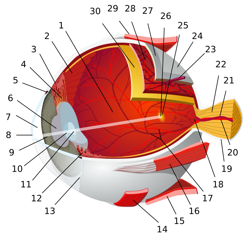
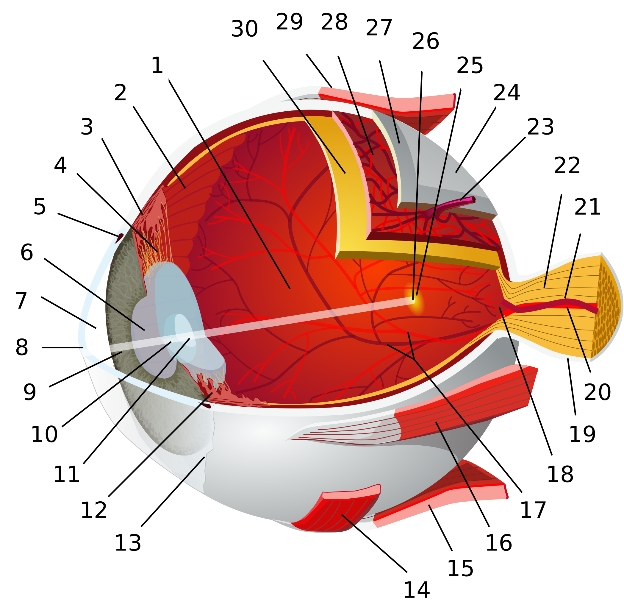
- posterior segment
- ora serrata
- ciliary muscle
- ciliary zonules
- Schlemm's canal
- pupil
- anterior chamber
- cornea
- iris
- lens cortex
- lens nucleus
- ciliary process
- conjunctiva
- inferior oblique muscle
- inferior rectus muscle
- medial rectus muscle
- retinal arteries and veins
- optic disc
- dura mater
- central retinal artery
- central retinal vein
- optic nerve
- vorticose vein
- bulbar sheath
- macula
- fovea
- sclera
- choroid
- superior rectus muscle
- retina

|

This image was selected as picture of the day on Wikimedia Commons for 20 June 2007. It was captioned as follows: English: A diagram of the human eye. Other languages:
Afrikaans: Diagram van die menseoog Čeština: Schematický průřez lidským okem Deutsch: Schematische Darstellung des menschlichen Auges English: A diagram of the human eye. Español: Diagrama de un ojo humano. Esperanto: Skema prezentado de la homa okulo Français : Schéma de l'œil humain. Italiano: Diagramma schematico dell'occhio umano. Magyar: Emberi szem szemléltető rajza Nederlands: Schematisch diagram van het menselijk oog Polski: Budowa gałki ocznej człowieka Português: Um diagrama de um olho humano. Română: Diagramă a ochiului uman. Slovenčina: Schematický prierez ľudským okom. Svenska: De olika delarna i ett mänskligt öga. Türkçe: İnsan gözü diyagramı. Беларуская: Схема будовы чалавечага вока. Русский: Схема глаза человека. 日本語: ヒトの眼球の構造図 中文: 人类眼球结构示意图 中文(繁體): 人類眼球結構示意圖 עברית : תרשים של העין האנושית. العربية : صورة تخطيطية للعين البشرية. |
Summary
| Description |
Afrikaans: 1: Agterste voorportaal 2: Getande rand 3: Siliêre spier 4: Siliêre sonule 5: Schlemm se kanaal 6: Pupil 7: Voorkamer 8: Kornea 9: Iris 10: Lenskorteks 11: Lenskern 12: Siliêre apparaat 13: Konjunktiva 14: Onderste skuinsspier 15: Onderste rektusspier 16: Mediale rektusspier 17: Retinale arteries en are 18: Optiese skyf 19: Dura mater 20: Sentrale retinale arterie 21: Sentrale retinale aar 22: Optiese senuwee 23: Vortikose aar 24: Bulba-skede 25: Makula 26: Put 27: Sklera 28: Choroïed 29: Boonste rektusspiere 30: Retina
العربية: 1. الغرفة الخلفية 2. الحاشية المشرشرة 3. العضلة الهدبية 4. النطيقة الهدبية 5. قناة شليم 6. البؤبؤ (حدقة العين) 7. الغرفة الأمامية 8. القرنية 9. القزحية 10. قشرة العدسة 11. نواة العدسة 12. النواتئ الهدبية 13. الملتحمة 14. العضلة المائلة السفلية 15. العضلة المستقيمة السفلية 16. العضلة المستقيمة الإنسية (الداخلية) 17. شرايين وأوردة الشبكية 18. القرص البصري 19. الأم الجافية 20. شريان الشبكية المركزي 21. وريد الشبكية المركزي 22. العصب البصري 23. الوريد الدواري 24. غمد المقلة 25. البقعة 26. النقرة 27. الصلبة 28. المشيمية 29. العضلة المائلة العلوية 30. الشبكية
Dansk: 1. Glaslegemet 2. Ora serrata 3. Akkomodationsmusklen (musculus ciliaris) 4. Zonulatråde 5. Schlemm'sk kanal 6. Pupil 7. Forkammeret 8. Hornhinden 9. Regnbuehinden 10. Linsebark 11. Linsekerne 12. Processus Ciliaris 13. Bindehinde (Conjunctiva) 14. Musculus obliquus inferior 15. Musculus rectus inferior 16. Musculus rectus medialis 17. Nethindens vener og arterier 18. Den blinde plet 19. Den hårde hjernehinde (Dura mater) 20. Centrale nethindearterie 21. Centrale nethindevene 22. Synsnerven 23. Vena vorticosum 24. Vagina bulbi 25. Den gule plet 26. Fovea centralis 27. Senehinden 28. Årehinden 29. Musculus rectus superior 30. Nethinden
Deutsch: 1:Glaskörper 2:Ora serrata 3:Corpus ciliare 4:Zonulafasern 5:Schlemm-Kanal 6:Pupille 7:vordere Augenkammer 8:Cornea 9:Iris 10:Linsenrinde 11:Linsenkern 12:Processus ciliares 13:Bindehaut 14:Musculus obliquus inferior 15:Musculus rectus inferior 16:Musculus rectus medialis 17:Gefäße der Netzhaut 18:Blinder Fleck 19:Hirnhaut 20:Arteria centralis retinae 21:Vena centralis retinae 22:Sehnerv 23:Vena vorticosa 24:Tenon-Kapsel 25:Gelber Fleck 26:Fovea centralis 27:Sclera 28:Choroidea 29:Musculus rectus superior 30:Retina
English: 1:posterior segment of eyeball 2:ora serrata 3:ciliary muscle 4:ciliary zonules 5:canal of Schlemm 6:pupil 7:anterior chamber 8:cornea 9:iris 10:lens cortex 11:lens nucleus 12:ciliary process 13:conjunctiva 14:inferior oblique muscle 15:inferior rectus muscle 16:medial rectus muscle 17:retinal arteries and veins 18:optic disc 19:dura mater 20:central retinal artery 21:central retinal vein 22:optic nerve 23:vorticose vein 24:bulbar sheath 25:macula 26:fovea 27:sclera 28:choroid 29:superior rectus muscle 30:retina
Español: Diagrama de un ojo humano. 1:humor vítreo 2:ora serrata 3:músculo ciliar 4:ligamento suspensorio del cristalino 5:canal de Schlemm 6:pupila 7:cámara anterior 8:córnea 9:iris 10:cortex del cristalino 11:núcleo del cristalino 12:cuerpo ciliar 13:conjuntiva 14:músculo oblícuo inferior 15:músculo recto inferior 16:músculo recto medial 17:arterias y venas retinianas 18:papila (punto ciego) 19:duramadre 20:arteria central retiniana 21:vena central retiniana 22:nervio óptico 23:vena vorticosa 24:conjuntiva bulbar 25:mácula 26:fóvea 27:esclerótica 28:coroides 29:músculo recto superior 30:retina
فارسی: ۱. زجاجیه ۲. حاشیه دندانهدار ۳. ماهیچه مژکی ۴. گردالکهای مژگانی ۵. مجرای اشلک ۶. مردمک ۷. اتاق جلویی ۸. قرنیه ۹. عنبیه ۱۰. عدسی ۱۱. عدسی ۱۲. زوائد مژگانی ۱۳. ملتحمه ۱۴. ماهیچه مایل زیرین ۱۵. ماهیچه راست زیرین ۱۶. ماهیچه راست میانی ۱۷. شبکیه ۱۸. صفحه بینایی ۱۹. سختشامه ۲۰. سرخرگ مرکزی شبکیه ۲۱. سیاهرگ مرکزی شبکیه ۲۲. عصب بینایی ۲۳. سیاهرگ حلقوی ۲۴. غلاف پیازی ۲۵. لکه زرد ۲۶. گودی مرکزی ۲۷. صلبیه ۲۸. مشیمیه ۲۹. ماهیچه راست بالایی ۳۰. شبکیه
Français : 1. Chambre postérieure (remplie d'humeur vitrée), 2. ora serrata, 3. muscle cilliaire, 4. ligament suspenseur, 5. canal de Schlemm, 6. pupille, 7. chambre antérieure (remplie d'Humeur aqueuse), 8. cornée, 9. iris, 10. cristallin cortical, 11. noyau du cristallin, 12. corps cilliaire, 13. conjonctive, 14. muscle oblique inférieur, 15. muscle droit inférieur, 16. muscle droit médian, 17. veines et artères rétinales, 18. papille optique ou point aveugle, 19. lame criblée, 20. artère centrale de la rétine, 21. veine centrale de la rétine, 22. nerf optique, 23. Veine vortiqueuse, 24. tissu conjonctif, 25. macula, 26. Fovéa, 27. sclère, 28. choroïde, 29. muscle droit supérieur, 30. rétine.
Hrvatski: 1. Stražnja očna sobica, 2. Nazubljena crta (ora serrata), 3. Cilijarni mišić, 4. Zonularna vlakna, 5. Schlemmov kanal, 6. Zjenica 7. Prednja očna sobica, 8. Rožnica, 9. Šarenica, 10. Kora (korteks) leće, 11. Jezgra leće, 12. Cilijarni nastavci, 13. Spojnica, 14. Donji kosi mišić, 15. Donji pravi mišić, 16. Medijalni pravi mišić, 17. Mrežnične arterije i vene, 18. Optički disk, 19. Tvrda ovojnica, 20. Središnja mrežnična arterija, 21. Središnja mrežnična vena, 22. Vidni živac, 23. Vrtložasta vena, 24. Ovojnica očne jabučice 25. Žuta pjega (macula lutea) 26. Fovea, 27. Bjeloočnica 28. Žilnica 29. Gornji pravi mišić, 30. Mrežnica.
Íslenska: 1. afturhólf, 2. laufarönd, 3. brárvöðvi, 4. brárgjörð, 5. blástokkur hvítu, 6. sjáaldur, ljósop, 7. framhólf, 8. glæra, hornhimna, 9. lithimna, lita, regnbogahimna, 10., 11. augasteinskjarni, 12. brárklakkar, 13. tára, augnslímhúð, 14. neðri skávöðvi, 15. neðri beinn, 16. miðlægur beinn, 17. sjónuslagæðir og sjónubláæðir, 18. sjóntaugardoppa, 19. heilabast, 20. sjónumiðjuslagæð, 21. miðjubláæð sjónu, 22. sjóntaug, 23. sveipbláæðar, 24. augnknattarslíður, 25. depill, 26. sjónugróf, 27. hvíta, augnhvíta, 28. æða, æðahimna, 29. efri beinn, 30. sjóna, nethimna, sjónhimna
日本語: 1:硝子体 2:鋸状縁 3:毛様(体)筋 4:チン小帯(毛様小帯) 5:シュレム管(強膜静脈洞) 6:瞳孔 7:前眼房(前房) 8:角膜 9:虹彩 10:水晶体皮質 11:水晶体核 12:毛様体突起 13:結膜 14:下斜筋 15:下直筋 16:内側直筋 17:網膜動脈・静脈 18:視神経乳頭 19:硬膜 20:網膜中心動脈 21:網膜中心静脈 22:視神経 23:渦静脈 24:テノン嚢 25:黄斑 26:中心窩 27:強膜 28:脈絡膜 29:上直筋 30:網膜.
ქართული: 0:ადამიანის თვალის ანატომია 1:თვალის უკანა სეგმენტი 2:არეა სერატა 3:ცილიალური სხეული 4:ცილიალური სარტყელი 5:შლემის არხი 6:გუგა 7:წინა საკანი 8:რქოვანა 9:ფერადი გარსი 10:ბროლის ქერქი 11:ბროლის ბირთვი| 12::ცილიალური სხეულის მორჩები 13:კონიუნქტივა| 14:ქვედა ირიბი კუნთი 15:ქვედა სწორი კუნთი 16:შიგნითა სწორი კუნთი 17:ბადურის არტერიები და ვენები 18:ბრმა ხალი(მხედველობის ნერვის დვრილი) 19:ტვინის მაგარი გარსი 20:ბადურის ცენტრალური არტერია 21:ბადურის ცენტრალური ვენა 22:მხედველობის ნერვი 23:გამჭოლი ვენა 24:ტუნიკა ბულბი 25:მაკულა 26:ფოვეა 27:სკლერა 28:უვეა 29:ზედა სწორი კუნთი 30:ბადურა
Македонски: 1:задна комора 2:ora serrata 3:цилијарен мускул 4:цилијарни зонули 5:Шлемов канал 6:пупила 7:предна комора 8:cornea 9:iris 10:lens cortex 11:lens nucleus 12:цилијарен процес 13:conjuntiva 14:инфериорен обличен мускул 15:инфериорен ректусен мускул 16:медијален ректусен мускул 17:ретинални артерии и вени 18:оптички диск 19:dura mater 20:централна ретинална артерија 21:централна ретинална вена 22:оптички нерв 23:вортикозна вена 24:топчест слој 25:macula 26:fovea 27:sclera 28:choroid 29:горен ректусен мускул 30:retina
Nederlands: 1:achterste oogkamer 2: ora serrata 3: musculus ciliaris 4: zonula ciliaris 5: kanaal van Schlemm 6: pupil 7: voorste oogkamer 8: hoornvlies 9: iris 10: lenskapsel 11: lenskern 12: processus ciliares 13: conjunctiva 14: m. obliquus inferior 15: musculus rectus inferior 16: musculus rectus medialis 17: retinale arteriën en venen 18: blinde vlek 19: hard hersenvlies 20: arteria centralis retinae 21: vena centralis retinae 22: nervus opticus 23: venae vorticosae 24: harde oogrok 25: gele vlek 26: fovea centralis 27: sclera 28: vaatvlies 29: m. obliquus superior 30: netvlies
Русский: 0:глаз 1:Задний сегмент глаза 2:зубчатый край 3:ресничная мышца 4:ресничный поясок 5:Шлеммов канал 6:зрачок 7:передняя камера 8:роговица 9:радужная оболочка 10:кора хрусталика 11:ядро хрусталика 12:цилиарный отросток 13:конъюнктива 14:нижняя косая мышца 15:нижняя прямая мышца 16:медиальная прямая мышца 17:артерии и вены сетчатки 18:слепое пятно (сосочек зрительного нерва) 19:твердая мозговая оболочка 20:центральная артерия сетчатки 21:центральная вена сетчатки 22:зрительный нерв 23:вортикозная вена 24:влагалище глазного яблока 25:жёлтое пятно 26:центральная ямка 27:склера 28:сосудистая оболочка глаза 29:верхняя прямая мышца 30:сетчатка
Slovenščina: 1. Zadajšnji očesni prekat 2. Nazobčana črta 3. Ciliarna mišica 4. Zinnova zonula 5. Schlemmov kanal 6. Zenica 7. Sprednji očesni prekat 8. Roženica 9. Šarenica 10. Skorja leče 11. Jedro leče 12. Grebeni ciliarnika 13. Veznica 14. Spodnja poševna mišica 15. Spodnja prema mišica 16. Medialna prema mišica 17. Arterije in vene mrežnice 18. Izstopišče vidnega živca 19. Trda možganska ovojnica (dura) 20. Centralna mrežnična arterija 21. Centralna mrežnična vena 22. Vidni živec 23. Vena vortikola 24. Tenonova ovojnica 25. Makula 26. Fovea 27. Beločnica 28. Žilnica 29. Zgornja prema mišica 30. Mrežnica
Polski: 1: komora tylna oka 2: rąbek zębaty siatkówki 3: mięsień rzęskowy 4: obwódka rzęskowa 5: kanał Schlemma 6: źrenica 7: komora przednia oka 8: rogówka 9: tęczówka 10: kora soczewki 11: jądro soczewki 12: wyrostek rzęskowy 13: spojówka 14: mięsień skośny, dolny 15: mięsień prosty, dolny 16: mięsień prosty, przyśrodkowy 17: tętnice i żyły siatkówki 18: tarcza nerwu wzrokowego 19: opona twarda 20: tętnica środkowa siatkówki 21: żyła środkowa siatkówki 22: nerw wzrokowy 23: żyła wirowata 24: otoczka gałki ocznej 25: plamka żółta 26: dołek centralny siatkówki 27: twardówka 28: naczyniówka 29: mięsień prosty, górny 30: siatkówka
Svenska: 1. glaskroppen 2. Ora serrata 3. ciliarmuskeln 4. Zonula ciliaris (kallas ibland zonula-trådar) 5. Schlemms kanal 6. pupill 7. främre ögonkammaren 8. hornhinna (kallas ibland kornea, cornea) 9. iris 10. lins (yttre skikt) 11. lins (kärna) 12. strålkropp 13. bindhinna (kallas ibland konjunktiva) 14. M. obliquus inferior 15. M. rectus inferior 16. M. rectus medialis 17. artärer och vener i näthinnan 18. synnervspapillen 19. hårda hjärnhinnan 20. Arteria centralis retinae 21. Vena centralis retinae 22. synnerven 23. (en av flera) Venae vorticosae 24. Vagina bulbi 25. gula fläcken (kallas ibland makula) 26. centralgropen (kallas ibland fovea) 27. senhinnan (kallas ibland sklera) 28. åderhinnan (kallas ibland koroidea, choroidea) 29. M. rectus superior 30. näthinnan (kallas ibland retina)
Türkçe: 1. arka oda 2.ora serrata 3.silier kası 4.kirpiksi bölge 5.Schlemm kanalı 6.göz bebeği 7.ön oda 8.kornea 9.iris 10.lens korteksi 11.lens çekirdeği 12.silier cisim (kirpiksi cisim) 13.konjunktiva 14.alt oblik kası 15.alt rektus kası 16.medial rektus kası 17.retinal arter ve venler 18. Izstopišče vidnega živca 19.dura mater 20.santral retinal arter 21.santral retinal ven 22.optik sinir 23.vortikoz veni (göz koroid veni) 24.Tenon kapsülü 25.makula 26.fovea 27.sklera 28.koroid (damar tabaka) 29.süperior rektus kası 30.retina
Українська: 1:задня камера ока 2:зубчастий край 3:циліарний м'яз 4:циннова зв'язка 5:шлеммів канал 6:зіниця 7:передня камера ока 8:рогівка 9:райдужна оболонка 10:капсула кришталика 11:ядро кришталика 12:циліарні відростки 13:кон'юнктива 14:нижній косий м'яз 15:нижній прямий м'яз 16:медіальний прямий м'яз 17:артерії і вени сітківки 18:сліпа пляма 19:тверда мозкова оболонка 20:центральна артерія сітківки 21:центральна вена сітківки 22:зоровий нерв 23:вортикозна вена 24:піхва очного яблука 25:жовта пляма 26:центральна ямка 27:склера 28:судинна оболонка ока 29:верхній прямий м'яз 30:сітківка
|
||||||||
| Date | |||||||||
| Source | References: [1] [2] [3] among others | ||||||||
| Author | Chabacano | ||||||||
| Permission (Reusing this file) |
I, the copyright holder of this work, hereby publish it under the following licenses:
This file is licensed under the Creative Commons Attribution-Share Alike 2.5 Generic, 2.0 Generic and 1.0 Generic license.
You may select the license of your choice. |
||||||||
| Other versions |
|
||||||||
| SVG development | This vector image was created with Inkscape, or with something else. |


![[1]](http://www.eyedesignbook.com/ch3/fig3-57bBG.jpg)
![[2]](http://www.eyedesignbook.com/ch3/fig3-57cBG.jpg)
![[3]](http://img225.imageshack.us/img225/3433/eye2ka5.png)
File history
Click on a date/time to view the file as it appeared at that time.
| Date/Time | Thumbnail | Dimensions | User | Comment | |
|---|---|---|---|---|---|
| current | 02:28, 25 February 2013 | | 1,237 × 1,208 (130 KB) | Ysangkok | scour |
| 13:07, 10 March 2007 |  | 1,237 × 1,208 (445 KB) | Ignacio Icke | trying to solve the centering issue | |
| 10:37, 8 March 2007 |  | 1,237 × 1,208 (403 KB) | Ignacio Icke | == Summary == {{Information |Description={{en|1:posterior chamber 2:ora serrata 3:ciliary muscle 4:ciliary zonules 5:canal of Schlemm 6:pupil 7:anterior chamber 8:cornea 9:iris 10:lens cortex 11:lens nucleus 12:ciliary process 13:conjuntiva 14:inferior ob |
File usage
More than 100 pages use this file. The following list shows the first 100 pages that use this file only. A full list is available.
- Amacrine cell
- Anterior chamber of eyeball
- Anterior segment of eyeball
- Aqueous humour
- Blood–ocular barrier
- Bowman's layer
- Bruch's membrane
- Capillary lamina of choroid
- Capsule of lens
- Choroid
- Ciliary body
- Ciliary muscle
- Ciliary processes
- Common tendinous ring
- Cone cell
- Cornea
- Corneal endothelium
- Corneal epithelium
- Corneal limbus
- Descemet's membrane
- Episcleral layer
- External limiting membrane
- Eye
- Fibrous tunic of eyeball
- Fovea centralis
- Fundus (eye)
- Ganglion cell layer
- Giant retinal ganglion cells
- Human eye
- Inner nuclear layer
- Inner plexiform layer
- Internal limiting membrane
- Intrinsically photosensitive retinal ganglion cell
- Iris (anatomy)
- Iris dilator muscle
- Iris sphincter muscle
- Layer of rods and cones
- Lens (vertebrate anatomy)
- Macula
- Mammalian eye
- Müller glia
- Night vision
- Optic cup (anatomical)
- Optic disc
- Optic pit
- Optics
- Ora serrata
- Outer nuclear layer
- Outer plexiform layer
- Pars plana
- Photoreceptor cell
- Posterior chamber of eyeball
- Posterior segment of eyeball
- Pupil
- Recurrent corneal erosion
- Red-eye effect
- Retina
- Retina bipolar cell
- Retina horizontal cell
- Retinal ganglion cell
- Retinal nerve fiber layer
- Retinal pigment epithelium
- Rod cell
- Sattler's layer
- Schlemm's canal
- Schwalbe's line
- Sclera
- Sensory nervous system
- Stroma of cornea
- Stroma of iris
- Suprachoroid lamina
- Tapetum lucidum
- Trabecular meshwork
- Uvea
- Visual processing
- Visual system
- Vitreous body
- Vitreous chamber
- Zonule of Zinn
- Talk:Photoreceptor cell/July 2006
- User:Fuelbottle/footers
- User:Madhero88/Cardiologytaskforce
- User:Passargea/Favourite pictures/Science
- User talk:Arad
- User talk:Ignacio Icke
- Wikipedia:Featured picture candidates/Eye-diagram no circles border.svg
- Wikipedia:Featured picture candidates/March-2007
- Wikipedia:Featured pictures/Sciences/Biology
- Wikipedia:Featured pictures thumbs/06
- Wikipedia:Picture of the day/July 2007
- Wikipedia:Templates for deletion/Log/2007 August 29
- Wikipedia:Wikipedia Signpost/2007-03-20/Features and admins
- Wikipedia:Wikipedia Signpost/2007-03-20/SPV
- Wikipedia:Wikipedia Signpost/2009-01-24/Dispatches
- Wikipedia talk:Picture of the day/Archive 3
- Wikipedia talk:Wikipedia Signpost/2009-01-24/Dispatches
- File:Eye-diagram no circles border.svg
- Template:Eye anatomy
- Template:Eye diagram
- Template:Eye diagram noframe
View more links to this file.
Global file usage
The following other wikis use this file:
- Usage on af.wikipedia.org
- Usage on ar.wikipedia.org
- لون
- اعتلال الشبكية عند الأطفال الخدج
- قرنية
- شبكية
- بوابة:طب/صورة مختارة
- ملتحمة
- قزحية
- حدقة
- عنبية (عين)
- أم جافية
- خلية غبارية
- خلية مخروطية
- خلية عصوية
- خلط مائي
- عدسة (تشريح)
- عضلة مستقيمة علوية
- عضلة مستقيمة سفلية
- بقعة الشبكية
- تكيف (عين)
- مشيمية
- خطأ انكسار
- عين الإنسان
- أمراض العين الفيروسية
- شريان شبكي مركزي
- صلبة (عين)
- خلية عصبية ثنائية القطب
- وريد شبكي مركزي
- ويكيبيديا:صور مختارة/علوم/علم الأحياء
- قالب:عين الإنسان
- خلية مستقبلة للضوء
- عصبون الشبكية
- نقرة مركزية
- خلية أماكرين
- عضلة مصرة القزحية
- ظهارة القرنية
- ظاهر الصلبة
- حوف القرنية (تشريح)
- غرفة العين الأمامية
- قطعة العين الأمامية
- قالب:عين الإنسان بلا إطار
- جسم زجاجي
- بوابة:تشريح
- بوابة:تشريح/صورة مختارة
- بوابة:تشريح/صورة مختارة/3
- جسم هدبي
- طبقة دوا
- غرفة العين الخلفية
- قالب:تشريح العين
View more global usage of this file.