| Anterior lacrimal crest | |
|---|---|
| Details | |
| Part of | maxilla |
| System | skeletal |
| Identifiers | |
| Latin | crista lacrimalis anterior |
| TA98 | A02.1.12.025 |
| TA2 | 782 |
| FMA | 75059 |
| Anatomical terms of bone | |
The anterior lacrimal crest is a bony projection on the frontal process of the maxilla. It creates the lateral margin of the lacrimal sac fossa and is continuous with the orbital margin. The medial palpebral ligament is attached to anterior lacrimal crest. It is an important structure to avoid damaging during rhinoplasty.
Structure[edit]
The anterior lacrimal crest is a bony projection on the frontal process of the maxilla in the skull.[1] It reaches the junction between the maxilla and the lacrimal bone.[2]
At its junction with the orbital surface is a small tubercle, the lacrimal tubercle, which serves as a guide to the position of the lacrimal sac.
The anterior lacrimal crest is much thicker and stronger than the posterior lacrimal crest.[3] It is one of the thickest parts of the orbit.[4] It is nearly always quite prominent, whilst the posterior lacrimal crest may be less prominent in some people.[5]
Relations[edit]
The lacrimal sac is directly behind the anterior lacrimal crest, which protects it as part of the fossa for the lacrimal sac.[6]
Function[edit]
The anterior lacrimal crest is the site of insertion of the medial palpebral ligament.[1] Some consider this a tendon of the orbicularis oris muscle.[1] The anterior lacrimal crest also protects the lacrimal sac.[6]
Clinical significance[edit]
Avulsion[edit]
The anterior lacrimal crest may be vulnerable to an avulsion fracture due to its connection to the medial palpebral ligament.[4]
Rhinoplasty[edit]
The anterior lacrimal crest is vulnerable to damage during osteotomy performed during rhinoplasty, a common plastic surgery.[2][7] It lies approximately 5 mm from the osteotomy incision line.[7]
See also[edit]
Additional images[edit]
-
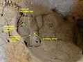 Cranium. Anterior lacrimal crest.Lacrimal bone.

References[edit]
![]() This article incorporates text in the public domain from page 161 of the 20th edition of Gray's Anatomy (1918)
This article incorporates text in the public domain from page 161 of the 20th edition of Gray's Anatomy (1918)
- ^ a b c Ritleng, P.; Bourgeon, A.; Richelme, H. (1983-03-01). "New concepts of the anatomy of the lacrimal apparatus". Anatomia Clinica. 5 (1): 29–34. doi:10.1007/BF01798870. ISSN 1279-8517. S2CID 931641.
- ^ a b Sachs, Michael Evan (1984). "Lacrimal system injury secondary to cosmetic rhinoplasty". Ophthalmic Plastic and Reconstructive Surgery. 3: 301–305. CiteSeerX 10.1.1.604.3718.
- ^ Tao, Hai; Ma, Zhi-zhong; Wu, Hai-Yang; Wang, Peng; Han, Cui (April 2014). "Anatomic study of the lacrimal fossa and lacrimal pathway for bypass surgery with autogenous tissue grafting". Indian Journal of Ophthalmology. 62 (4): 419–423. doi:10.4103/0301-4738.121137. ISSN 0301-4738. PMC 4064215. PMID 24817745.
- ^ a b Stranc, M. F. (1970). "The pattern of lacrimal injuries in naso-ethmoid fractures". British Journal of Plastic Surgery. 23 (4): 339–346. doi:10.1016/S0007-1226(70)80072-8. ISSN 0007-1226. PMID 5475760.
- ^ Bisaria, K K; Saxena, R C; Bisaria, S D; Lakhtakia, P K; Agarwal, A K; Premsagar, I C (October 1989). "The lacrimal fossa in Indians". Journal of Anatomy. 166: 265–268. ISSN 0021-8782. PMC 1256759. PMID 2621144.
- ^ a b Merbs, Shannath; DeParis, Sarah (2020-01-01), Dorafshar, Amir H.; Rodriguez, Eduardo D.; Manson, Paul N. (eds.), "3.4 - Ocular Considerations: Ectropion, Entropion, Blink, Ptosis, Epiphora", Facial Trauma Surgery, London: Elsevier, pp. 367–378, ISBN 978-0-323-49755-8, retrieved 2021-09-23
- ^ a b Sarıaltın, Yücel; Ortak, Turgut; Öz, Cem; Kuş, Emel; Yunusoğlu, Gülşah Uslu; Doğan, Şevket; Çetin, Meltem (2019-10-01). "Radiological assessment of the lateral osteotomy line–lacrimal system distance on three-dimensional models". Journal of Cranio-Maxillofacial Surgery. 47 (10): 1608–1616. doi:10.1016/j.jcms.2019.07.026. ISSN 1010-5182. PMID 31400844. S2CID 199539798.