Original file (935 × 323 pixels, file size: 126 KB, MIME type: image/jpeg)
Summary

|
File:Lungvolumes.svg is a vector version of this file. It should be used in place of this JPG file.
File:LungVolume.jpg → File:Lungvolumes.svg
For more information, see Help:SVG. |
 |
| Description |
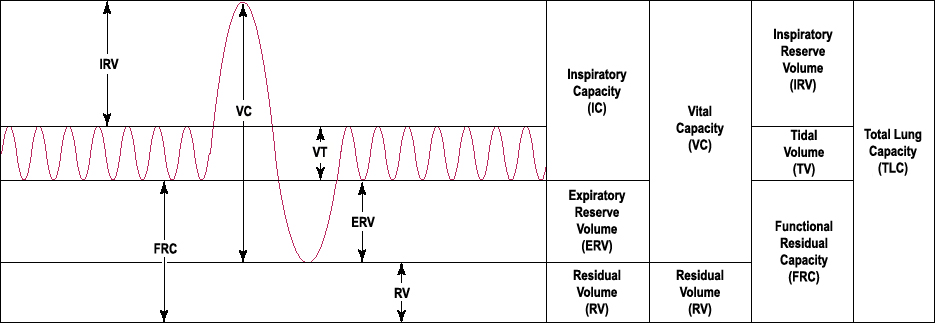
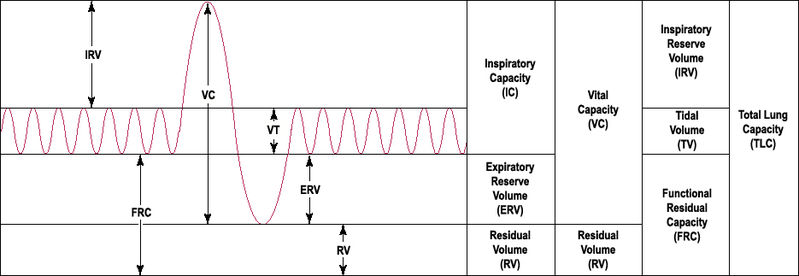
English: This is a prototypical output of a 'en:spirometer'. The vertical axis signifies the en:volume and the horizontal axis signifies en:time. The bottom left corner equals zero lung volume at the start of the spirometer recording session. The first small en:amplitude part of the en:sinusoid depicts repeated resting state involuntary breathing. The amplitude of this small sinusoid corresponds to the 'Tidal Volume'. The large positive amplitude spike represents voluntary inspiration to maximal volume or 'Total Lung Volume'. The large negative amplitude spike represents forced expiration to the lowest possible en:physiological lung volume called 'Residual Volume'.
All of the en:lung volumes commonly referred to in the literature are labeled. Vihsadas 21:31, 18 April 2007 (UTC) Vihsadas (talk) 05:52, 7 March 2008 (UTC) Vihsadas 20:10, 14 April 2007 (UTC) |
| Date | 13 April 2007 (original upload date) |
| Source | Transferred from en.wikipedia |
| Author | Original uploader was Vihsadas at en.wikipedia |
| Permission (Reusing this file) |
Released into the public domain (by the author). |
| Other versions | Derivative works of this file: LungVolume cs.png |
Licensing
| This work has been released into the public domain by its author, Vihsadas. This applies worldwide. In some countries this may not be legally possible; if so: |
Being somewhat adept at "nit-picking," I noted that the graphic on Lung Volume shows "VT" for Tidal Volume. Based on what I observe in the rest of the graphic, that graphic element should be, "TV."
Certainly, this is not a grievous error.
Original upload log
- 2007-04-18 21:43 Vihsadas 935×323×8 (128701 bytes) This is a prototypical output of a 'spirometer'. The y-axis signifies the volume, with the bottom left corner equaling 'zero volume'. The sinusoid comes from repeated resting state breathing (small amplitude sinusoid) 'Tidal Volume', one inspiratory segme
- 2007-04-18 21:29 Vihsadas 935×323×8 (128531 bytes) '
- 2007-04-13 01:06 Vihsadas 836×327×8 (116712 bytes) This image was UPDATED using the original image that was posted here. Therefore it is fair usage. There is no copyright holder. The original owner of the image made the file and uploaded it here, I updated it with new information. Since it was already upl
File history
Click on a date/time to view the file as it appeared at that time.
| Date/Time | Thumbnail | Dimensions | User | Comment | |
|---|---|---|---|---|---|
| current | 12:51, 1 June 2008 | 935 × 323 (126 KB) | Mrug | {{Information |Description={{en|This is a prototypical output of a 'en:spirometer'. The vertical axis signifies the en:volume and the horizontal axis signifies en:time. The bottom left corner equals zero lung volume at the start of the spir |
File usage
Global file usage
The following other wikis use this file:
- Usage on ar.wikipedia.org
- Usage on de.wikipedia.org
- Usage on es.wikipedia.org
- Usage on id.wikipedia.org
- Usage on it.wikipedia.org
- Usage on ja.wikipedia.org
- Usage on ml.wikipedia.org
- Usage on nl.wikipedia.org
- Usage on sr.wikipedia.org
- Usage on sv.wikipedia.org